Beam Delivery System
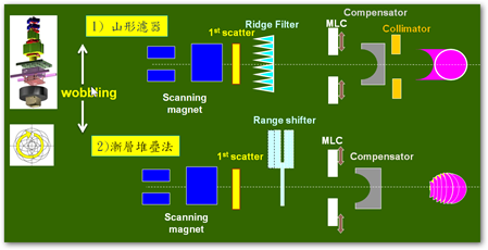
The proton therapy machine has two different treatment techniques to meet the
individual needs of tumor treatment, Pencil Beam Scanning and Wobbling. The
principle of Wobbling involves sending a narrow proton beam through a magnetic
field of the X- and Y-axes and deflectors while it expands with a rotation of a specific
radius into a broader beam with a wider range. When it passes through the ridge
filter, the vertical axis expands into SOBP, passes through the compensator and
collimator, and then conforms before reaching the body. Another method that can
reduce unnecessary normal tissue dose involves using layer stacking and a multileaf
collimator (MLC) that can adjust the field size. This way, the dose distribution can be
done as shown in the figure below. The figure clearly shows that the orange block,
which represents normal tissue, receives a much smaller dose. This advanced
technology is being used by our Proton Therapy Center. Since the energy output of
the cyclotron is fixed (230MeV), by using the energy selection system (ESS), the
energy level that reaches the treatment site can be adjusted. Different energy levels
can penetrate different depths. Therefore, by using adjustment mechanisms, the
tumor is divided into different layers of the same width. Treatment starts from the
deepest layer and gradually moves upwards, while the use of the multileaf
collimator follows the 3D shape of the tumor.

The principle of Pencil Beam Scanning involves sending a narrow proton beam
through a magnetic field of the X- and Y-axes and distributing it directly in the field
by scanning. This technique controls the weighted dose of each point in the field to
achieve "Intensity Modulated Proton Therapy (IMPT)". This advanced technology is
currently in the Proton Therapy Optimization Plan and is essentially the direction of
the future of proton therapy.

Patient Position System
Digital Imaging Positioning System:
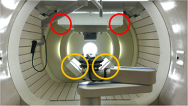
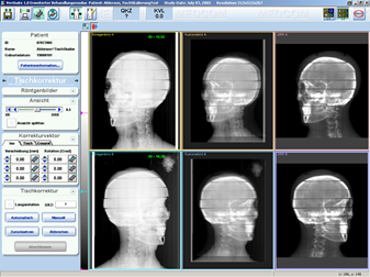
To ensure the positional accuracy of proton therapy, every treatment room is
equipped with a digital imaging guiding system, which acquires images through
three different methods: digital radiography, fluoroscopy and computed
tomography.
A. Digital radiography can acquire two orthogonal two-dimensional
images to match the treatment site.
B. Fluoroscopy can view lesion displacement caused by a variety of
reasons, such as breathing, and adjust treatment to such breathing with
the aid of proton therapy equipment.
C. Computed tomography is used to compare its image with the simulated image from
the computer treatment plan system to ensure the accuracy of the treatment site.

X-ray Imaging Positioning System

Digital Radiography
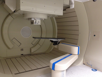
Robotic Couch: after the Digital Imaging Positioning System determines the
correct patient position on the couch, this information is immediately sent to the
robotic couch for a 6-dimensional shift. The robotic couch is a specialized couch
developed by combining an industrial computer-controlled robotic arm and a
radiotherapy couch. The robotic couch has a total of six drive motors, which enable
it to move along six axes and rotate with an accuracy of 0.01 mm. Not only can it
measure the weight of a patient (maximum user weight 200 kg), but it can also
compensate for inclinations due to weight. It is the most advanced and accurate
couch among radiotherapy equipment today.
