Traditionally, physicians need to increase the safety margin around the tumor for conventional radiation therapy to ensure that the radiation remains within the tumor area; thus, we cannot effectively spare normal tissue organ. These limitations were mainly due to patient setup uncertainties and internal organ motion. Image-guided radiation therapy (IGRT) provides high-resolution, two-dimensional imaging and three- dimensional volumetric images prior to treatment to pinpoint bony landmarks or internal markers to ensure treatment accuracy and that the same position is targeted in every treatment session. Furthermore, it minimizes the deviation of internal organ motion or patient setup uncertainties, as well as decreases the irradiation to normal organs.
Through more precise targeting of the beam to tumors, doses to the
tumors can be increased and the non-target volumes can be reduced.
Higher radiation doses in tumors have been shown to enhance tumor
control and better spare non-target regions, reducing the possibility of side
effects after radiotherapy.
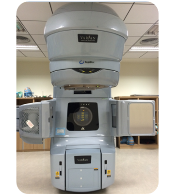
The On-Board Imager® kV imaging system with cone-beam CT and imaging
fusion technique provides more precise localization information and better
treatment quality and accuracy. Due to the need of x-ray imaging and
patient positioning correction before each treatment for image guided
radiotherapy, it takes about four to five minutes more than the
conventional radiation therapy treatment time.
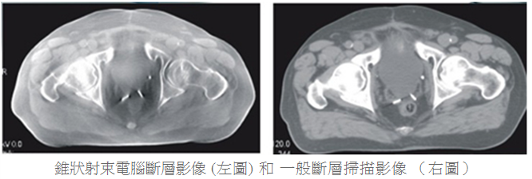
Cone beam CT (left) vs. regular CT images (right)