|
顯微鏡核心實驗室
MICROSCOPY CORE LABORATORY
|
|
|
實 驗 室 設 備 |
|
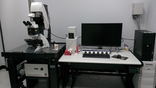
正立白光雷射共軛焦顯微鏡 (TCS SP8X)
 |
。研究發表
。設備聯絡人
姓名:陳志俊
黃美綾
梅秀富
電話:(03)3281200
分機:7706 |
- 技術原理
- 儀器規格
- 機型:Leica TCS SP8X
- 光源:
- Visible laser
- White light laser system:
pulsed laser
- super
continuous exciting range: 470nm-670nm
- 1nm adjustable
- >1.5mW for each wavelength
- UV laser
- 顯微鏡:LEICA DM6000 CS 顯微鏡 壹台
- 物鏡:
- Plan APO 10x, dry, NA 0.40, WD≧2.2mm
- Plan APO 20x, NA 0.70, coverglass, WD≧0.59mm
- Plan APO 40x, oil, NA 1.30, WD≧0.24mm
- Plan APO 63x, oil, NA 1.40, WD≧0.14mm
- Plan APO 100x, oil, NA 1.40, WD≧0.09mm
- APO 10x UVI, water immersion, NA 0.30, LWD≧3.6mm
- APO 20x UVI, water immersion, NA 0.50, LWD≧3.5mm
- APO 40x UVI, water immersion, NA 0.80, LWD≧3.3mm
- 掃瞄器:
- for UV/405 nm-VIS-IR-gSTED
with 4 independent laser ports
- Light gate FLIM technology
- Dual scan Tandem scanner
- High resolution scan mode:
- Scan field of view(FOV) at least 22 mm, speed 7fps@512x512 in FOV 22mm, 84fps @ 512 x 16
- Max. scan resolution 8192 x 8192, 16 bits grey scale,
digitalization 12bits or 18 bits.
- High speed scan mode:
- Resonent scanner 12kHz (bi-direction 24kHz) , speed
40fps @ 512x512, 420fps @ 512 x 16
- 24kHz max. scan resolution 832 x 832, 16 bits grey
scale, digitalization 12bits or 18 bits
- CCD:
- 1.4 Mpixel (1392 x 1040)
- Exposure time 4us-10min
- 影像擷取形式:
◆ Bright field
◆ Fluorescence
◆ Reflection
◆ Normaski differential interference contrast (DIC)
- Build-in Scanner' confocal detector:
Multi-band
Spectrophotometer(400-850 nm)with 4PMT tube (2 cooling hybrid GaAsP/APD detectors + 2 cooled PMT detectors)
- 軟體:
- Leica Application Suite X
- Imaris 3D/4D real time image software
- Metamorph image analysis
software
|
|
|