
Page 26 - 分子轉譯影像中心季刊XI
P. 26
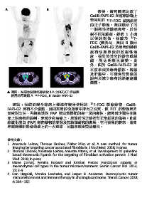
A                              B
                                                                           最  後  ,研究團隊比較了
                                                                     Ga68-FAPI-02 與臨床診斷上
                                                                     常用到的         18 F-FDG 做癌細胞

                                                                     的正子影像。圖四顯示了同
                                                                     一個惡性肺腺癌患者,於注
                                                                     射不同示蹤劑,經過 1 小時
                                                                     以  後   的  影  像  。 相較      於    18 F-
                                                                     FDG (圖四A),圖四 B 顯示
                                                                     Ga68-FAPI-02 於患者的轉移
                                                                     處 有 明 顯 累 積 的 放 射 性 活
                                                                     度,相對於其它的器官或組

                                                                     織 , 對 比 明 顯 且 清 楚 。 此
                                                                     外,由於 Ga68-FAPI-02 並
                                                                     不會堆積於患者腦部、脾臟
                                                                     及肝臟中,可避免對影像的
                                                                     誤判及減少患者的輻射劑量
                                                                     暴露。


         圖四、為惡性肺腺癌患者於 1 h 之PET/CT 掃描圖,
                           18
         使用的示踨劑 A.  F-FDG , B. Ga68-FAPI-02

          結論:相較於現今臨床上標準作業所使用的                                        18 F-FDG 影像藥物,Ga68-
      FAPI-02 具有不少優點,例如其易於從血液中排出之性質,使 PET 的影像具有
      高度對比性,再結合其對 FAP 蛋白質標靶的專一及內噬性,能夠減少醫生在臨

      床上對患者旳誤判,亦減少在治療上,具放射性之藥物對正常細胞的傷害。此纖
      維激化蛋白 (FAP) 導向藥物的開發及其轉譯研究的進展,在可預見的將來,將會
      是腫瘤標靶影像領域上的一大突破,且臨床運用效益無限。


       參考文獻:
       1. Anastasia Loktev, Thomas Lindner, Walter Mier, et al. A new method for tumor
          imaging by targeting cancer associated fibroblasts. J Nucl Med. 2018; in press
       2. Thomas Lindner, Anastasia Loktev, Annette Altann, et al. Development of quionline
          based theranostic ligands for the targeting of fibroblast activation protein. J Nucl
          Med. 2018; in press
       3. Eliane Cortez, Pernilla Roswall and Kristian Pietras. Functional subsets of
          mesenchymal cell types in the tumor microenvironment. Semin Cancer Biol. 2014;
          25: 3-9.
       4. Dan Høgdall, Monika Lewinska, and Jesper B. Andersen. Desmoplastic tumor
          microenvironment and immunotherapy in cholangiocarcinoma. Trends Cancer. 2018;
          4: 239 - 255