|
|
|
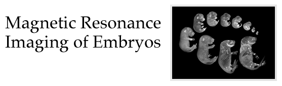
Magnetic resonance imaging (MRI)
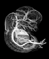
is used to non-destructively investigate the internal and external anatomy of embryos. This technique produces three-dimensional image arrays that can be volume-rendered and it allows the specimen to be dissected electronically to reveal structures of interest. These pages provide many sample images and movies of embryos, all acquired by MRI. There are also explanations of the techniques used to produce the images and movies, and information about how to order "The Digital Atlas of Mouse Embryology", a CD containing over two thousand images, and fifty quicktime movies.
Sample Images and Movies of Animal Embryos The Multi-dimensional Human Embryo CD of Mouse Embryology by MRI How MRI of Embryos is Performed Collaborators' Pages (password-protected) Research Conducted by Brad Smith The Center for In Vivo Microscopy Links to related sites Contact Information for Comments and Questions |
| Animal Embryos | Human Embryos | Embryo CD | About MRI | Collaborators | Brad's Research | CIVM | Links | Contact Info |
For information:
brdsmith@umich.edu
Bradley R. Smith
Director, Medical and Biological Illustration
University of Michigan
Ann Arbor, Michigan
USA
©1997 Bradley R. Smith