|
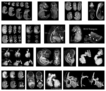
Mouse, chick, and opposum embryos
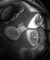
have been studied by magnetic resonance imaging (MRI). The sample pictures on this page demonstrate the level of anatomical detail in the images produced by MRI. Because MRI can produce three-dimensional image data non-destructively, the intact embryos can be volume-rendered to provide whole-specimen views, single-slice views, and cut-away views from any orientation. Sample pictures also show how special MRI contrast materials can be injected into the vasculature of the embryos to highlight the blood vessels and developing heart.
|


