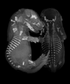

| Bradley R. Smith, Ph.D. Associate Professor and Director Medical and Biological Illustration School of Art and Design Associate Professor, Radiology University of Michigan Ann Arbor, Michigan 48109 USA Cardiovascular Development Developmental morphology of the cardiovascular system and the association of vascular anomalies with other organ system dysmorphogenesis. Magnetic Resonance Microscopy Embryo Culture Systems Development of embryo culture chambers for imaging ex-utero live mouse embryos while inside the magnetic resonance scanner. Magnetic Resonance Contrast Agents Development of magnetic resonance contrast materials to emphasize the location and timing of developmental events. Embryo Imaging Development of perfusion techniques, coil designs, imaging schemes, data analysis and image rendering for improved magnetic resonance study of embryos. Magnetic Resonance Microscopy of Human Embryos (The Multidimensional Human Embryo) Dale S. Huff, G. Allan Johnson, Adrianne Noe The need for information on human embryos to better understand processes of development and disease, and the difficulty in obtaining well-documented and well-preserved specimens presents an important challenge to understanding normal and abnormal development. With funding from the National Institute of Child Health and Human Development (NICHD) we finishing a program to generate a digital atlas of human embryology that is interactive and multidimensional. The atlas is based on magnetic resonance microscopy of human embryos that are already a part of the highly respected Carnegie Collection of Human Embryos. This digital atlas goes far beyond any material that is currently available to study human embryology by providing a complete three-dimensional data set for each of 18 human embryos representing Carnegie stages 10 through 23, a critical embryonic time period for organogenesis. It will greatly facilitate the work of clinicians, investigators, and students of human development by bringing diverse pieces of information together in an easily retrievable and widely available form, replacing a process that traditionally takes days or weeks of library research to perform. In-vitro MR Microscopy of Mouse Embryos North Carolina Biotechnology Center The mouse embryo is one of the most common embryos used by pharmaceutical developers, toxicologists, and researchers to study developmental biology. Mammalian embryos are also the most difficult embryos to observe during critical stages of development because un-like other vertebrate and in-vertebrate embryos, the mammalian embryo cannot simply be flushed out of the uterus or opened up like an egg, and it is too small to be studied with standard X-rays or ultrasonography. The consequence is that most current technologies measure and observe sacrificed embryos (fixed specimens) rather than investigating the live embryo. Techniques are being developed to combine magnetic resonance microscopy and whole embryo culture techniques to make possible the investigation of live mouse embryos. Magnetic Resonance Microscopy of Mouse Embryo Specimens (The Digital Atlas of Mouse Embryology) North Carolina Biotechnology Center Elwood Linney, G.Allan Johnson A multidimensional, digital atlas has been completed that contains magnetic resonance images of normal mouse embryos from 9.5 days after conception to the newborn. The images include dramatic surface views as well as cross-sectional views from the transverse, coronal, and sagittal planes for each embryo. Several movies have also been included to demonstrate growth of the embryos and to present a variety of visualization tools available for studying and documenting embryonic anatomy. These images were acquired and organized into this atlas to facilitate the otherwise very tedious and difficult process of learning embryology, and to arouse interest in the important topic of developmental biology. These images are also organized as a reference for researchers who want to understand better the embryological anatomy of their own specimens and to understand how their images relate to the whole embryo at many stages of development. Publications Smith, B.R. Visualizing human embryos. Scientific American, 280:76-81 (March), 1999. Dewhirst MW.; Ong, ET.; Braun, RD.; Smith, BR.; Klitzman, B.; Evans, SM.; Wilson, D. Quantification of Longitudinal Tissue pO2 Gradients in Window Chamber Tumors: Impact on Tumor Hypoxia. British Journal of Cancer, 79:1717-1722, 1999. Smith, BR.; Huff, DS.; Johnson GA. Magnetic resonance imaging of embryos: An internet resource for the study of embryonic development. Computerized Medical Imaging and Graphics, 23:33-40, 1999. Huang, GY; Wessels, A; Smith, BR; Linask, KK; Ewart, JL; Lo, C. Alteration in Connexion 43 Gap Junction Gene Dosage Impairs Conotruncal Heart Development. Developmental Biology, 198:32-44, 1998. Smith, BR; Shattuch, MD; Hedlund, LW; and Johnson, GA. Time-course imaging of rat embryos in utero with magnetic resonance microscopy. Magnetic Resonance in Medicine, 39:673-677,1998. Smith, BR. Magnetic resonance microscopy with cardiovascular applications. Trends in Cardiovascular Medicine, 6:247-254, 1996. Smith, BR.; Linney, E.; Huff, DS.; Johnson, GA. Magnetic resonance microscopy of embryos. Computerized Medical Imaging and Graphics, 20:483-490, 1996. Jaber, M.; Koch, JW.; Rockman, HA.; Smith, B.; Bond, RA.; Sulik, K.; Ross, J. Jr.; Lefkowitz, RJ.; Caron, MG.; Giros, B. Essential role of beta-adrenergic receptor kinase 1 in cardiac development and function. Proc Natl Acad Sci USA, 93:12974-12979, 1996. Smith, BR., Johnson, GA., Groman, EV., Linney, E. Magnetic resonance microscopy of mouse embryos. Proceedings of the National Academy of Science., USA, 91:3530-3533, 1994. Mellin, AF., Cofer, GP., Smith, BR., Suddarth, SA., Hedlund, LW., and Johnson, GA. Three dimensional magnetic resonance microangiography of rat neurovasculature. Magnetic Resonance in Medicine, 32:199-205, 1994. Johnson, GA., Benveniste, H., Black, RD., Hedlund, LW., Maronpot, RR., Smith, BR. Histology by Magnetic Resonance Microscopy. Magnetic Resonance Quarterly, 9:1-30, 1993. Smith, BR, Effmann, EL, Johnson, GA., MR microscopy of Chick Embryo Vasculature. Journal of Magnetic Resonance Imaging, 2:237-240, 1992. Effmann, EL., Johnson, GA., Smith, BR., Talbot, GA., Cofer, G. Magnetic resonance microscopy of chick embryos in ovo. Teratology, 38:59-65, 1988. Effmann, EL., Whitman, SA., Smith, BR. Aortic arch development. Radiographics, 6:1065 1089, 1986. |
| Home | Animal Embryos | Human Embryos | Embryo CD | About MRI | Collaborators | CIVM | Links | Contact Info |