
Page 64 - 林口醫研部 3月份電子報
P. 64
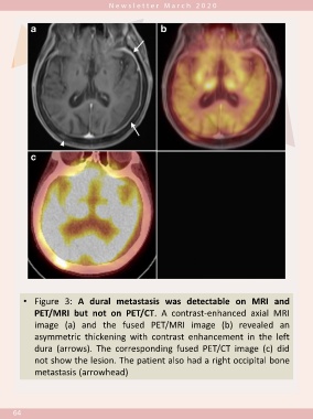
• Figure 3: A dural metastasis was detectable on MRI and
               PET/MRI but not on PET/CT. A contrast-enhanced axial MRI


               image (a) and the fused PET/MRI image (b) revealed an
               asymmetric thickening with contrast enhancement in the left


               dura (arrows). The corresponding fused PET/CT image (c) did
               not show the lesion. The patient also had a right occipital bone


               metastasis (arrowhead)


      64