
Page 63 - 林口醫研部 3月份電子報
P. 63
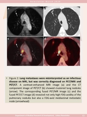
• Figure 2: Lung metastases were misinterpreted as an infectious

               disease on MRI, but was correctly diagnosed on PET/MRI and

               PET/CT. A contrast-enhanced MRI image (a) and the CT

               component image of PET/CT (b) showed clustered lung nodules

               (arrow). The corresponding fused PET/MR image (c) and the

               fused PET/CT image (d) revealed not only high FDG-avidity of the

               pulmonary nodules but also a FDG-avid mediastinal metastatic

               node (arrowhead)


                       Department of Medical Research & Development,  Linkou Newsletter                                   63