| Instruments |
|
|
Laparoscopic Inguino-Femoral Hernia Repair
|
|
| This technique is identical to the TAPP technique. However, it entirely takes place in the preperitoneal space. Thus, it is a totally extraperitoneal repair. The indications, the operating room setup and the placement of the trocars are also identical to the setup described in the new TAPP Repair. |
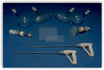
Instruments
- 2 USSC VERSAPORT* trocar-5 mm
- 1 USSC ENDO SHEARS* Instrument with cautery connection
- 1 USSC ENDO DISSECT* Instrument
- 1 reusable Storz 5 mm dolphin nose grasper
- 1 6" x 6" USSC SURGIPRO* Mesh (Open weave) or a Mesh
- 1 disposable USSC PROTACK* (30 tacks) or 5mm USSC™ Tacker.
- 1 TEP USSC Disposable Set ( 1 distension balloon trocar, 1 structural trocar, 1 pump)
Entry and creation of the pre-peritoneal space
No pneumoperitoneum is created. Instead, a small incision is made below the umbilicus (midline). The midline is exposed. An incision is made slightly lateral to the midline aponevrosis ( on the ipsilateral side of the inguinal hernia). The anterior and posterior Rectus Muscle Sheaths are exposed. A small space is developed bluntly with a finger. The balloon expander is then inserted. The remainder of the technique varies with the instrument used.
Creation of the pre-peritoneal space
The USSC™ Preperitoneal distension balloon system is inserted in the pre-peritoneal space. The balloon is inflated under direct vision with the telescope in place.
Once the balloon has been inflated to full capacity, it is deflated and the entire system removed.
The USSC™ Structural Balloon Trocar is inserted into the pre-peritoneal space. The structural balloon is inflated through the balloon inflation port with 4 pumps of the bulb. (CAUTION: do not overinflate.)
By depressing the latch of the exterior sponge-seal, the Structural Balloon Trocar will be locked in position. This device will seal off the pre-peritoneal space and maintain the trocar in place.
| The repair |
|
The extra-peritoneal or pro-peritoneal space is now well-expanded and ready for the inguinal hernia repair. The two lateral 5 mm trocars are inserted under direct vision. They can be placed laterally or in midline position. The operator should be certain to enter the pre-peritoneal space and not the intra-abdominal cavity to avoid a CO2 leak. This will generate a pneumo-peritoneum and will greatly impair visualization.
As for the TAPP repair, the anatomy has to be clearly identified. Cooper's ligament should be first visualized as well as the Inferior Epigastric Vessels. The direct peritoneal sac is easy to identify and reduce. An indirect hernia sac should be bluntly pulled away from the Spermatic Cord and the inguinal canal using the atraumatic grasper. The hernia sac should be dissected as medially as possible to allow the Mesh to cover the entire inguinal region.
The Mesh is inserted via the medial, sub-umbilical trocar and deployed over the entire inguinal region. A 6" x 6" SURGIPRO MESH is used. The Protack* Instrument is used to anchor the mesh (Cooper's Ligament, around the epigastric vessels, lateral to the inguinal ring, midline rectus Musle Aponevrosis).
Once the repair is completed, the pre-peritoenal space is deflated and the trocars removed. The facial defect is closed.
| Technical Notes |
|
References: