Chang Gung Memorial Hospital
GB BIG-5 home
Home People Facility Research Collaboration resources
 
>>  Electron
 Microscopy
bar
>>  Confocal  Microscopy
bar
  • Confocal Microscopy
upright confocal  

Upright Confocal Microscopy

Model: Leica TCS SP2
Microscope: Leica DMRE HC(UV)
Lasers:
 Visible laser
  ◆Ar laser (458, 476, 488 and 514nm)
  ◆He-Ne laser (543nm)
  ◆He-Ne laser (633nm)
 UV laser
  ◆Ar-UV laser
 Scanner
  ◆“k” Scanner with 3-individual fiber-
   coupling laser ports and optics
Detector: Multi-band Spectrophotometer
     (400-850nm) with 4PMT tube
Software: Leica confocal software
Image Forms:
 ◆Bright field
 ◆Fluorescence
 ◆Reflection
 ◆Phase contrast
 ◆Normaski differential interference contrast
     (DIC)

     
inverted confocal  

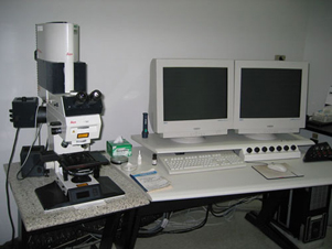
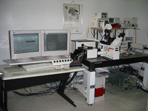
Inverted Confocal Microscopy

Model: Leica TCS SP2
Microscope: Leica DM IRBE HC(UV)
Lasers:
 Visible laser
  ◆Ar laser (458, 476, 488 and 514nm)
  ◆He-Ne laser (543nm)
  ◆He-Ne laser (633nm)
 IR laser
  ◆Spectra-physics' Tsunami Solid-State
      Mode-locked Ti: Sapphire laser
  ◆Tenability: 690 ~ 1080 nm
  ◆Laser pulse type: Pico second mirror set
Scanner:
  ◆“k” Scanner with 3-individual fiber-
   coupling laser ports and optics
Detector: Multi-band Spectrophotometer
     (400-850nm) with 3PMT tube
Software: Leica confocal software
Image Forms:
 ◆Bright field
 ◆Fluorescence
 ◆Reflection
 ◆Phase contrast


            TEL:+886-3-3281200 ext. 8174  
            E-MAIL:c12037@adm.cgmh.org.tw  
            ADD:No.5, Fusing St., Gueishan Township, Taoyuan County 333, Taiwan (R.O.C.)