What are Obstetric Ultrasound Scans?
Obstetric Ultrasound is the use of ultrasound scans in pregnancy. Since its introduction in the late 1950’s ultrasonography has become a very useful diagnostic tool in Obstetrics. Currently used equipments are known as real-time scanners, with which a continous picture of the moving fetus can be depicted on a monitor screen. Very high frequency sound waves of between 3.5 to 7.0 megahertz (i.e. 3.5 to 7 million cycles per second) are generally used for this purpose. They are emitted from a transducer which is placed in contact with the maternal abdomen, and is moved to "look at" (likened to a light shined from a torch) any particular content of the uterus. Repetitive arrays of ultrasound beams scan the fetus in thin slices and are reflected back onto the same transducer. The information obtained from different reflections are recomposed back into a picture on the monitor screen (a sonogram, or ultrasonogram). Movements such as fetal heart beat and malformations in the feus can be assessed and measurements can be made accurately on the images displayed on the screen. Such measurements form the cornerstone in the assessment of gestational age, size and growth in the fetus.

Why and when is Ultrasound used in Pregnancy?
Ultrasound scan is currently considered to be a safe, non-invasive, accurate and cost-effective investigation in the fetus. It has progressively become an indispensible obstetric tool and plays an important role in the care of every pregnant woman.
The main use of ultrasonography are in the following areas:
1. Diagnosis and assessment of early pregnancy.
The gestational sac can be visualized as early as four and a half weeks of gestation and the yolk sac at about five weeks.
2. Threatened miscarriage.
The viability of the fetus can be documented in the presence of vaginal bleeding in early pregnancy. Fetal heart motion is usually clearly depictable by 7 weeks. If this is observed, the probability of a continued pregnancy is greater than 97 percent. Missed abortion and blighted ovum will usually give typical pictures of a deformed gestational sac and absence of fetal poles or heart beat. Ultrasonography is also indispensible in the early diagnosis of ectopic pregnancies and molar pregnancies.
3. Determination of gestational age and assessment of fetal size.
Fetal body measurements reflect the gestational age of the fetus. This is particularly true in early gestation. In patients with uncertain last menstrual periods, such measurements must be made as early as possible in pregnancy to arrive at a correct dating for the patient. In the latter part of pregnancy measuring body parameters will allow assessment of the size and growth of the fetus and will greatly assist in the diagnosis and management of intrauterine growth retardation (IUGR).
The following measurements are usually made:
|
a) The Crown-rump length (CRL) This measurement can be made between 7 to 13 weeks and gives very accurate estimation of the gestational age. Dating with the CRL can be within 3-4 days of the last menstrual period. |
|
|
b) The Biparietal diameter (BPD) The diameter between the 2 sides of the head. This is measured after 13 weeks. It increases from about 2.4 cm at 13 weeks to about 9.5 cm at term. Different babies of the same weight can have different head size, therefore dating in the later part of pregnancy is generally considered unreliable.
|
|
|
c) The Femur length (FL) Measures the longest bone in the body and reflects the longitudinal growth of the fetus. Its usefulness is similar to the BPD. It increases from about 1.5 cm at 14 weeks to about 7.8 cm at term. |
|
|
d) The Abdominal circumference (AC) The single most important measurement to make in late pregnancy. It reflects more of fetal size and weight rather than age. Serial measurements are useful in monitoring growth of the fetus. |
4. Placental localization.
Ultrasonography has become indispensible in the diagnosis or exclusion of placenta previa, and other placental abnormalities as in diabetes, fetal hydrops, Rh isoimmunization and severe intrauterine growth retardation .
5. Multiple pregnancies.
In this situation, ultrasonography is invaluable in determining the number of fetuses, the chorionicity, fetal presentations, evidence of growth retardation and fetal anomaly,the presence of placenta previa, and any suggestion of twin-to-twin transfusion.
6. Hydramnios and Oligohydramnios.
Excessive or decreased amount of amniotic fluid) can be clearly depicted by ultrasound. In both these situations, careful ultrasound examination should be made to exclude intrauterine growth retardation and congenital malformation in the fetus such as intestinal atresia, hydrops fetalis or renal dysplasia. .
7. Fetal malformation.
Many structural abnormalities in the fetus can be reliably diagnosed by an ultrasound scan, and these can usually be made before 20 weeks. Common examples include hydrocephalus, anencephaly, myelomeningocoele, achondroplasia and other dwarfism, spina bifida, exomphalos, duodenal atresia and fetal hydrops. With more recent equipments, conditions such as cleft lips/palate, congenital cardiac abnormalities and Down syndrome are more readily recognised. Markers for chromosomal abnormalities such as the fetal nuchal translucency (the area at the back of the neck) have also been defined to enable detection of these abnormal fetuses. Ultrasound can also assist in other diagnostic procedures in prenatal diagnosis such as amniocentesis, chorionic villus sampling, percutaneous umbilical blood sampling and in fetal therapy.
8. Other areas.
Ultrasonography is of great value in other obstetric conditions such as:
|
a) confirmation of intrauterine death. |
||
|
b) confirmation of fetal presentation in uncertain cases. |
||
|
c) evaluating fetal movements, tone and breathing in the Biophysical Profile. |
||
|
d) diagnosis of uterine and pelvic abnormalities during pregnancy e.g. fibromyomata and ovarian cyst. |
The Schedule
There is no hard and fast rule as to the number of scans a woman should have during her pregnancy. A scan is ordered when an abnormality is suspected on clinical grounds. Otherwise a scan is generally booked at about 7 weeks to confirm pregnancy, exclude ectopic or molar pregnancies, confirm cardiac pulsation and measure the crown-rump length for dating.
A second scan is performed at 18 to 20 weeks to look for congenital malformations, exclude multiple pregnancies and to verify dates and growth. Placental position is also determined.
A third scan may sometimes be done at around 34 weeks to evaluate fetal size and assess fetal growth. Placental position is verified.
Many centers are now doing a scan at around 13-14 weeks to measure the nuchal skin fold thickness for the purpose of evaluating the risk for Down Syndrome.
The total number of scans will vary depending on whether a previous scan has detected certain abnormalities that require follow-up assessment.
What is often referred to as a Level II scan merely indicates a "targeted" examination where it is done when an indication is present or when an abnormality is suspected in a previous examination. In fact professional bodies such as the American Institute of Ultrasound in Medicine does not endorse or encourage the use of these terms. A more "thorough" examination is usually done at an a perinatal center or specialised clinic where more expertise and better equipments may be present.
One should not dwell too much on the definitions or guildlines for a level II ultrasound scan. The sonologist should always try very hard to look for and assess any abnormality that may be present in the fetus. It is not very meaningful to be talking about level III or even level IV scans.
Whether a pregnancy must be scanned on a 'routine' basis at 18 to 20 weeks is still a matter of some controversy.
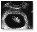
|
Fetus clearly visible within uterine cavity |
|
|

|
|
