Laparoscopic Hysterectomy-With and Without Salpingo-Oophorectomy
Revolutionary
advancements in endoscopy have dramatically changed
the practice of gynecology. With the introduction of the newest generation of endoscopic instruments, we have become more proficient in laparoscopically assisted hysterectomies with bilateral salpingo-oophorectomies.
There is still
controversy as to whether traditional vaginal hysterectomy is the better
procedure compared with its laparoscopic counterpart. Our experience with
laparoscopic hysterectomy demonstrated that the laparoscopic approach is
superior in most patients with myomas, previous
cesarean sections, undiagnosed endometriosis, chronic pelvic inflammatory
disease and unexpected intestinal pathology.
The greatest
advantage of a laparoscopic hysterectomy is the significant reduction in
postoperative pain, discomfort and the shorter recovery period. Compared to
traditional open surgical procedures, laparoscopic hysterectomy has reduced the
hospital stay and cost by
|
INSTRUMENTS |
Note: The telescope used in these cases is a Storz straightforward telescope 0
with parallel eyepiece and 5mm instrument channel. A Storz Video system is
used. All patients are given Toradol (Roche Pharmaceuticals)
30mg IM and Cefizox (Fujisawa
USA) 2 Gm IV during the induction phase.
|
THE PROCEDURE |
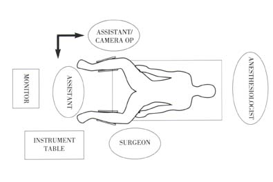
Operating Room
Setup
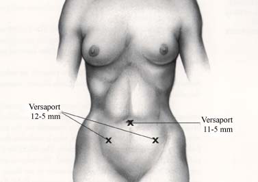
Trocar Placement
The
Technique
- STEP
The patient is
placed in lithotomy position with Allen universal
stirrups. A Kronner Manipujector
is inserted. The abdomen and perineum are prepped in the usual manner.
The pneumoperitoneum is created. The trocars
are inserted. The intraabdominal cavity is
visualized. Using the uterine manipulator, the uterus and adnexa
are moved, and all aspects of the internal genitalia are exposed and examined.
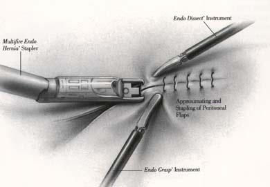
If intraabdominal adhesions are present, an enterolysis will be performed using the ENDO SHEARS* and
ENDO DISSECT* instruments connected to the electrocautery.

- STEP
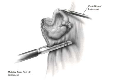
The ovary is
grasped at its ligament and elevated with an ENDO DISSECT* instrument. The infundibulopelvic ligament is identified and spread. A Multifire ENDO GIA*
The same procedure
is repeated on the other side.
- STEP
The operative site
is brought into telescopic view by moving the Kronner
Manipujector. The anterolateral
aspect of the uterovesical junction is placed under
tension. Using the ENDO SHEARS* instrument connected to an electrocautery
source, the peritoneal bladder flap is developed in traditional fashion.

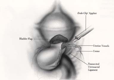
The dissection is
started laterally and continued anteriorly around the
cervix. The bladder flap is gently peeled away with the ENDO SHEARS*
instrument. The hemostasis is controlled with electrocautery. The posterior sheath of the broad ligament
is dissected down to the uterosacral ligament. The uterosacral ligaments are transected using an ENDO SHEARS*
instrument.
- STEP
The uterine vessels
(artery and vein) are identified. At this level, they usually run parallel to
the uterus. The ureter will also be lateral to the
vessels and also parallel to the uterus. Stay as close as
possible to the uterus while clipping, almost clipping the vessels into the
cervix.
The assistant
places a curved sponge stick into the posterior vagina.
- STEP
A posterior colpotomy is made with the ENDO SHEARS* instrument. This
vaginal incision is not made by cutting but merely by using the electrocautery power with the ENDO SHEARS* instrument to
transect. It is performed slowly so that the pneumoperitoneum
will not be rapidly lost (CO

The vaginal will be
packed and sealed with wet Kerlix gauze. The pneumoperitoneum is recreated. The colpotomy
is completed anteriorly and laterally. The colpotomy is extended around the base of the cervix on the
vagina until the uterus is detached from the vagina.
- STEP
If a bilateral oophorectomy is to be performed, a Multifire
ENDO GIA
The vaginal pack is
removed. A large Kocher clamp is passed transvaginally into the intraabdominal
cavity under direct vision. The uterus and both tubo-ovarian
complexes are removed via the colpotomy.
- STEP
The colpotomy is then closed transvaginally
with interrupted size
The abdomen is
irrigated with Ringer¡¦s lactate solution; desufflated
and all the cannulas are removed. The
References:
- Summitt RL et al: Randomized comparison of
laparoscopic assisted hysterectomy with standard vaginal hystectomy in an outpatient setting (
- Nezhat F et al: Laparoscopic versus abdominal
hysterectomy. (