Surgical Management of Ectopic Pregnancy
|
|
INTRODUCTION |
|
In the not-so-distant past (
The advent of these two tests is the major reason that
maternal morbidity is now measured per
History of the Procedure: Prior to
In
Following Tait's accomplishment, little occurred in the
management of ectopic pregnancy until the
Pregnancy tests in the
Problem: An ectopic pregnancy occurs outside of the uterus.
Approximately
Of tubal pregnancies, the ampulla is the most common site
of implantation (
Frequency: The incidence of ectopic pregnancy most commonly is
reported as the number of ectopic pregnancies per
In
About
Etiology: The main cause of ectopic pregnancy is salpingitis.
The morphologic sequelae of acute salpingitis can be seen in approximately
Pathophysiology: Delay or prevention of passage
of the fertilized ovum (blastocyst) to the uterine cavity by the factors as
above or factors inherent in the embryo that result in premature implantation.
Clinical: The most common clinical presentation is pelvic
pain and/or vaginal spotting. This usually occurs
Pain usually is the result of stretching of the
peritoneum over the tube. Once the tube ruptures, pain usually decreases or
disappears.
If the tube has ruptured, the patient may present in
shock with tachycardia and hypotension. Shoulder pain from diaphragmatic
irritation is a late sign and seldom is seen today.
|
|
WORKUP |
|
Lab Studies:
- Human chorionic
gonadotropin (hCG) (quantitative)
- The
quantitative level of beta hCG found in ectopic pregnancy varies.
Serum-beta hCG levels correlate with the size and gestational age in
normal embryonic growth. In a normal pregnancy, the beta hCG level
doubles every
- The
discriminatory zone of beta hCG is the level above which a normal
intrauterine pregnancy (IUP) is reliably visualized. Once beta hCG has
reached a level of
- The lack of an
IUP when the beta is above the discriminatory zone represents an ectopic
pregnancy or a recent abortion.
- Perform serial
hemoglobin or hematocrit levels to quantify blood loss.
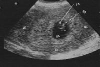
Imaging Studies:
- Endovaginal
ultrasonography can be performed in the outpatient clinic or emergency
room to exclude an IUP.
- Definite IUP: A
gestational sac with a sonolucent center (greater than
- Probable
abnormal IUP: A gestational sac larger than
- Definite
ectopic pregnancy: The presence of a thick, brightly echogenic, ringlike
structure located outside the uterus with a gestational sac containing an
obvious fetal pole, yolk sac, or both. This is an unusual finding.
- No definite IUP (empty uterus): An empty uterus on endovaginal
ultrasound in patients with a serum beta hCG greater than the
discriminatory cut-off value is an ectopic pregnancy until proven
otherwise. An empty uterus also may represent a recent abortion.
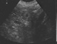
- Other
ultrasonographic findings include an adnexal mass(usually a corpus
luteum, occasionally hematoma), free cul-de-sac fluid, and/or severe
adnexal tenderness with probe palpation. Patients with no definite IUP
and the above-mentioned findings may be at high risk for an ectopic
pregnancy.
- An appreciation
for the sonographic spectrum of ultrasound findings in ectopic pregnancy
may allow physicians to recognize an early ectopic pregnancy. The spectrum
of sonographic findings in ectopic pregnancy include the following:
- Tubal ring: An
echogenic ringlike structure found outside of the uterus represents an
early ectopic pregnancy.
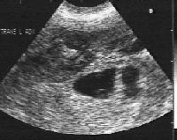
- Extrauterine
mass: The presence of a tender adnexal mass on ultrasound suggests an
ectopic pregnancy. One study suggested that the presence of any adnexal
mass other than a simple cyst was the most significant ultrasound finding
for the diagnosis of ectopic pregnancy.
- Interstitial
ectopic: An interstitial ectopic pregnancy is one that implants at the
highly vascular region of the uterus near the insertion of the fallopian
tube. These types can grow larger than those within the fallopian tube,
because the endometrial tissue is more expandable. Because of the
increased size and partial endometrial implantation, these advanced
ectopics can be misdiagnosed as an IUP. An aid in the diagnosis of an
interstitial ectopic is the eccentric location of the gestational sac. It
is important to evaluate the amount of uterine myometrium surrounding the
gestational sac and echogenic decidual layer. This is termed the
myometrial mantle. At least
- Heterotopic pregnancy: This is a combined IUP and ectopic pregnancy.
It may occur in approximately
- Extrauterine empty gestational sac: The presence of an extrauterine
mass with a thick, brightly echogenic band (ring) also may represent an
ectopic.
- Hemosalpinx: Fallopian tubes may fill with blood or free fluid.
- Ruptured
ectopic: Findings on ultrasound include free fluid or clotted blood in
the cul-de-sac or in the intraperitoneal gutters, such as Morison pouch.
Diagnostic Procedures:
- Culdocentesis
can be performed to diagnose blood in the cul-de-sac. Ultrasound is
relatively noninvasive and is more sensitive for cul-de-sac fluid but
cannot distinguish between peritoneal fluid and blood.
- Menstrual
aspiration can be performed and content sent for pathology to exclude a
missed or incomplete abortion.
|
|
TREATMENT |
|
Medical therapy: The greatest advance in the
management of ectopic pregnancy since Tait has been the development of medical
management. In certain carefully selected patients, IM methotrexate can be both
safe and effective therapy.
To determine acceptable candidates for methotrexate
therapy, first establish the diagnosis by one of the following criteria:
- Abnormal
doubling rate of beta hCG and sonographic identification of a gestational
sac outside of the uterus
- Abnormal
doubling rate of beta hCG, empty uterus, and menstrual aspiration with no
chorionic villi
Once the diagnosis is established, the following criteria
also should be met:
- Hemodynamically
stable
- Reliable,
compliant patient who will come back for follow-up care
- Ectopic
pregnancy less than
- Absence of fetal
cardiac activity on ultrasound
- No evidence of
tubal rupture
- Beta hCG less
than
Criteria
On the day of treatment, obtain CBC, creatinine, ALT,
AST, and beta hCG levels. Evidence of hepatic or renal compromise
contraindicates methotrexate therapy. Blood type, Rh, and antibody screen also
are obtained, and all Rh-negative patients are given Rh immunoglobulin. A
menstrual aspiration or D & C also should be performed and the sample
evaluated for the absence of chorionic villi. If villi are obtained, this is
evidence of an abortion and the patient is treated appropriately. If no villi
are found, then the patient is a candidate for methotrexate therapy. Persons
with experience at floating villi in saline and identifying them under a
dissecting microscope can use this procedure. Those without experience may need
to wait for histologic confirmation of the absence of villi.
Day
Methotrexate (
Day
Patient returns for beta hCG level. This level may be
higher than pretreatment level. Day
Day
Draw beta hCG, CBC, and AST levels. If beta hCG has
dropped
If beta hCG has not dropped at least
If the patient develops increasing abdominal pain after
methotrexate, repeat a transvaginal scan to evaluate for possible rupture.
Using this protocol, Stovall et al achieved a
Drug Category: Antineoplastics ?Inhibit cell
proliferation by destroying rapidly dividing cells.
|
Drug Name |
Methotrexate (Folex, Rheumatrex) ?Acts as a folate
antagonist. |
|
Adult Dose |
Ectopic pregnancy |
|
Pediatric Dose |
Not indicated |
|
Contraindications |
Documented hypersensitivity; caution in pregnancy;
caution in lactating patients; caution in those with alcohol abuse history;
caution in patients with liver dysfunction or infection; caution if patient
has impaired liver or renal function or bone marrow depression |
|
Interactions |
Combination of acitretin and methotrexate may increase
risk of hepatotoxicity; combination of aspirin and methotrexate may increase
methotrexate levels; combination of Cox |
|
Pregnancy |
D ?Unsafe in pregnancy |
|
Precautions |
Caution in those with alcohol abuse history; caution in
patients with liver dysfunction or infection; caution if patient has impaired
liver or renal function or bone marrow depression |
Surgical therapy: Surgical therapy may use either
the open laparotomy or laparoscopic route. The choice of route depends on the
skill of the surgeon, available equipment, and the condition of the patient.
For example, if at
Consider all of these factors when deciding on the best
possible route to approach an ectopic pregnancy.
Preoperative details: If the patient is
hemodynamically unstable, obtain large-bore venous access and start fluid
resuscitation. Do not delay the operation. The patient has an active bleeding
site, and it must be stopped as soon as possible.
Place a Foley catheter prior to starting the procedure.
Either a Hulka tenaculum or a HUMI device inserted into
the uterus may be helpful in manipulating the tube during surgery.
Intraoperative details:
Surgical
procedures
Regardless of the route of approach, Salpingectomy is
indicated in the following situations:
- The ectopic has
ruptured.
- Future fertility
is not desired.
- This is a
sterilization failure.
- It is a
previously reconstructed tube.
- Sterilization is
requested.
- Hemorrhage
continues after salpingotomy.
- The ectopic is
in the blind ending distal segment after a previous partial salpingectomy.
- This is a
chronic tubal pregnancy.
In the absence of any of the above indications for
salpingectomy, salpingotomy may be performed.
If the ectopic presents at the fimbria, then fimbrial
evacuation is feasible, in the absence of indications for salpingectomy.
Partial salpingectomy may be indicated if the pregnancy
is in the mid portion of the tube, none of the indications for salpingectomy
are present, and the patient may be a candidate for later tubal reanastomosis.
Laparoscopy
- Salpingectomy
technique
- Desiccate the tube between the uterus and the ectopic, utilizing
bipolar cautery.
- Compress and desiccate the tubo-ovarian artery, while preserving the
utero-ovarian artery and ligament.
- Cut along the desiccated path, closer to the specimen, leaving a
pedicle for hemostasis.
- Repeat until the tube is free and can be removed.
Salpingotomy technique
- Infiltrate the mesosalpinx with vasopressin (
- With the knife or needle electrode, make a
- Insert the aquadissector deep into the incision.
- Fluid from the aquadissector under pressure dissects and dislodges
the ectopic and clots.
- Irrigate the bed well.
- If trophoblastic tissue remains, the use of vasopressin may lead to
anoxia and death of the trophoblasts, preventing postoperative growth.
- Further dissection may damage the tube and usually is not performed.
- The products of conception are then removed through the
- If needed, products of conception can be reduced to smaller pieces
using biopsy forceps or the aquadissector.
- Bleeding may be controlled with pressure from grasping forceps for
- Arterial bleeding may require pinpoint bipolar desiccation.
- Diffuse venous bleeding is best controlled with monopolar current. A
spark or arc is created using a current of
- Uncontrollable bleeding may require application of an endo loop to
provide compression for
- If bleeding continues, suture of the mesosalpingeal vessels may be
attempted.
Fimbrial evacuation technique
- Grasp the fimbria and rotate to allow insertion of the
aquadissector.
- Fluid under pressure dissects and dislodges ectopic and clots.
- Remove the products of conception.
Partial salpingectomy technique
- Perform bipolar desiccation across the tube on both sides of the
ectopic.
- Divide the tube at the sites of desiccation.
- The mesosalpinx under the ectopic then can either be desiccated or
ligated with an endo loop.
- Remove the products of conception.
Laparotomy salpingectomy
technique
- Clamp the tube between the uterus and the ectopic, using a Pean or
similar clamp. Cut the pedicle free and ligate the pedicle with a suture
ligature.
- Clamp, cut, and ligate the tubo-ovarian artery, while preserving the
utero-ovarian artery and ligament.
- Continue to clamp cut and ligate the mesosalpinx until the tube is
free and can be removed.
Salpingotomy technique
- Infiltrate the mesosalpinx with vasopressin (
- With the knife or needle electrode, make a
- Insert the aquadissector, or a syringe filled with saline, deep into
the incision.
- Fluid from the aquadissector, or syringe, under pressure dissects
and dislodges the ectopic and clots.
- Irrigate the bed well.
- If trophoblastic tissue remains, the prior injection of vasopressin
may lead to anoxia and death of the trophoblasts, preventing
postoperative growth.
- Further dissection may damage the tube and usually is not performed.
- Bleeding may be controlled with pressure from blunt tissue forceps
for
- Arterial bleeding may require pinpoint bipolar desiccation.
- Diffuse venous bleeding is best controlled with monopolar current. A
spark or arc is created using a current of
- Uncontrollable bleeding may require application of suture ligature
to provide compression for
- If bleeding continues, suture of the mesosalpingeal vessels may be
attempted.
- The tubal incision is left open and not repaired.
Fimbrial evacuation technique
- Grasp the
fimbria and insert the aquadissector or a syringe filled with saline.
- Fluid under
pressure dissects and dislodges ectopic and clots.
- Remove the
products of conception.
Partial salpingectomy technique
- Place a clamp
through an avascular area in the mesosalpinx under the ectopic. This
creates a space through which
- Tie the free
ties around the tube on each side of the ectopic.
- Cut free and
remove the isolated portion of the tube containing the ectopic.
Postoperative details: Postoperatively, most patients
with an ectopic pregnancy are able to leave the hospital as soon as they have
left the recovery room.
In patients who were in shock or had to receive blood
transfusions, the postoperative observation should be longer and include
observation that the kidneys are functioning normally and the patient has
regained normal hemodynamics.
Follow-up care: All patients who have not had
the entire ectopic pregnancy removed by salpingectomy need to have their weekly
hCG levels observed until they return to nonpregnant levels. If during this
time span the hCG level either plateaus or rises, treat with methotrexate.
Patients should all be on some form of effective
contraception until such time as their hCG levels have returned to nonpregnant
levels.
|
|
COMPLICATIONS |
|
While not exactly a complication, cervical pregnancy
should be discussed. Cervical pregnancy is an ectopic that has implanted in the
cervix. This can cause severe hemorrhage if it starts to separate from the
cervix. There are few muscle fibers in the cervix, thus there is no
constriction around the hypertrophied blood vessels that developed for the
pregnancy. With no pressure on the vessels, profuse hemorrhage can occur.
In recent years, ultrasound diagnosis has improved to the
point where the diagnosis is made much more frequently in asymptomatic
patients. This leads to many more options in management.
Previously, the only treatment was surgical with curettage of the implantation
site. This frequently led to such profuse hemorrhage that surgeons recommended
having the patient's abdomen open with ligatures placed around the uterine
arteries or hypogastric arteries prior to starting the curettage. Hysterectomy
frequently was the result.
Currently, the recommended treatment is either
hysterectomy for those who do not desire fertility or methotrexate for those
who do desire fertility. Since patients who receive methotrexate occasionally
develop severe hemorrhage, observe these patients closely for
Medical pitfalls
Certain diagnostic pitfalls can occur for the physician sonographer
in the diagnosis of ectopic pregnancy.
- Low beta hCG
levels: Consider beta hCG levels carefully in conjunction with ultrasound
findings. Low beta hCG levels may be misleading. Kaplan et al found that
- Location of
gestational sac: An ectopic pregnancy may be mistaken for a hemorrhagic
corpus luteum cyst or bowel. Advanced ectopics are misdiagnosed as an IUP
when the gestational sac and contents have a normal appearance but the
sonographer overlooks the extrauterine position of the sac. Employing a
systematic approach utilizing the longitudinal and transverse image planes
of the uterus and adnexa is mandatory. The ultrasound examination is not
complete when an IUP is identified.
- Pseudogestational
sac: A pseudogestational sac can be confused with a gestational sac or
with embryonic demise. An ectopic pregnancy may stimulate the endometrium,
causing a fluid collection within the endometrium.
- Hemorrhage and
hypovolemic shock
- Infection
- Loss of
reproductive organs following surgery
- Infertility
- Urinary and/or
intestinal fistulas following complicated surgery
- Disseminated
intravascular coagulation
|
|
OUTCOME AND
PROGNOSIS |
|
The prognosis for patients with an ectopic pregnancy is
good for those with an early diagnosis.
Fertility may be conserved in those patients diagnosed
with an ectopic pregnancy. The earlier the diagnosis is made and treatment
administered, the higher the likelihood of subsequent fertility.
Thirty years ago, when the diagnosis was seldom made
prior to rupture, the likelihood of a subsequent normal healthy term pregnancy
was only about
|
|
PICTURES |
|
|
Caption:
Picture |
|
|
|
Picture Type: Photo |
|
Caption:
Picture |
|
|
|
Picture Type: Photo |
|
Caption:
Picture |
|
|
|
Picture Type: Photo |
|
|
BIBLIOGRAPHY |
|
- Abbott J, Emmans
LS, Lowenstein SR: Ectopic pregnancy: ten common pitfalls in diagnosis. Am
J Emerg Med
- Ackerman TE,
Levi CS, Dashefsky SM: Interstitial line: sonographic finding in
interstitial (cornual) ectopic pregnancy. Radiology
- Chandra L, Jain
A: Maternal serum creatine kinase as a biochemical marker of tubal
pregnancy. Int J Gynaecol Obstet
- Cosin JA, Bean
M, Grow D: The use of methotrexate and arterial embolization to avoid
surgery in a case of cervical pregnancy. Fertil Steril
- Emerson DS,
Cartier MS, Altieri LA: Diagnostic efficacy of endovaginal color Doppler
flow imaging in an ectopic pregnancy screening program. Radiology
- Graham H:
Eternal Eve. Eternal Eve
- Graham M,
Cooperberg PL: Ultrasound diagnosis of interstitial pregnancy: findings
and pitfalls. J Clin Ultrasound
- Jafri SZ,
Loginsky SJ, Bouffard JA: Sonographic detection of interstitial pregnancy.
J Clin Ultrasound
- Kadar N, DeVore
G, Romero R: Discriminatory hCG zone: its use in the sonographic
evaluation for ectopic pregnancy. Obstet Gynecol
- Koonin LM,
MacKay AP, Berg CJ: Pregnancy-related mortality surveillance--United
States,
- Lavie O, Beller
U, Neuman M: Maternal serum creatine kinase: a possible predictor of tubal
pregnancy [see comments]. Am J Obstet Gynecol
- Leach RE, Ory
SJ: Modern management of ectopic pregnancy [see comments]. J Reprod Med
- Mateer JR, Aiman
EJ, Brown MH: Ultrasonographic examination by emergency physicians of
patients at risk for ectopic pregnancy. Acad Emerg Med
- Mateer JR,
- Stovall TG,
Kellerman AL, Ling FW: Emergency department diagnosis of ectopic
pregnancy. Ann Emerg Med
- Stovall TG, Ling
FW, Cope BJ: Preventing ruptured ectopic pregnancy with a single serum
progesterone. Am J Obstet Gynecol
- Stovall TG, Ling
FW, Gray LA: Single-dose methotrexate for treatment of ectopic pregnancy
[see comments]. Obstet Gynecol