|
|
|
|
|
|
|
|
 |
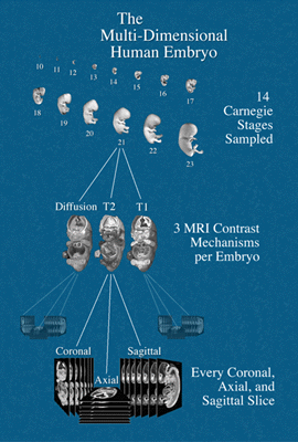
Summary Human embryo specimens representing 10 stages of develoment were imaged by magnetic resonance microscopy (MRM) to produce three-dimensional image data sets. Each embryo was imaged with three different magnetic resonance pulse sequences (T1-, T2-, and Diffusion-weighted) to generate three distinct types of pictures. A complete set of coronal, sagittal, and axial images were produced from each image data set. Several major organs were isolated from each T1-weighted embryo data set using image segmentation methods and separate image data sets were created to represent each of these organs. Additionally, each embryo was optically photographed under a low-power microscope. All of these images are available from this website. This work was carried out between July 1996 and June 2001 and was funded by the National Institute of Child Health and Human Development (NICHD) of the National Institutes of Health (NIH). MRM was performed at the Center for In-vivo Microscopy at Duke University. Image processing and data managment was performed at the School of Art and Design, University of Michigan. |
|
| Detailed Project Description | ||
| Overview Diagram | ||
| Collaborators | ||
| Technical Data | ||